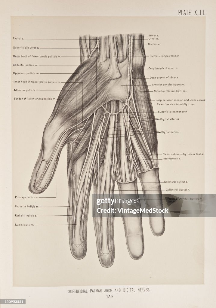
Superficial Palmar Arch & Digital Nerves
The superficial palmar arch is formed by the terminal part of the ulnar artery, and is completed by the superficials volae, or a branch from the radialis indicis or princeps pollicis, and sometimes, though rarely, by a large median artery, 1899. From 'The Treatise of the Human Anatomy and Its Applications to the Practice of Medicine and Surgery, Volume I' (1899). Digital nerves of ulnar nerve are branches on the dorsum of the hand. The dorsal branch of the ulnar nerve divides into two dorsal digital branches. (Photo by VintageMedStock/Getty Images)

EINE LIZENZ KAUFEN
Wie darf ich dieses Bild verwenden?
475,00 €
EUR
Bitte beachten Sie: Bilder, die historische Ereignisse darstellen, können Motive oder Beschreibungen beinhalten, die nicht der gegenwärtigen Auffassung entsprechen. Sie werden in einem historischen Kontext bereitgestellt. Weitere Informationen.
DETAILS
Einschränkungen:
Bei kommerzieller Verwendung sowie für verkaufsfördernde Zwecke kontaktieren Sie bitte Ihr lokales Büro.
Bildnachweis:
Redaktionell #:
150953551
Kollektion:
Archive Photos
Erstellt am:
1. Januar 1899
Hochgeladen am:
Lizenztyp:
Releaseangaben:
Kein Release verfügbar. Weitere Informationen
Quelle:
Archive Photos
Objektname:
T1676661_146
Max. Dateigröße:
2437 x 3477 px (20,63 x 29,44 cm) - 300 dpi - 4 MB